Long-tern use of Hydroxychloroquine

Blog vol 6.19. Long-tern use of Hydroxychloroquine.
Chloroquine, which was developed in the 1930s to replace quinine as the preventive of choice for malaria, is a great treatment for rheumatoid arthritis. My first introduction to the drug was in 1987, when I had to take it daily during an eye mission in Ecuador. I remember that it tasted terrible.
Chloroquine works against autoimmune diseases like rheumatoid arthritis and systemic lupus erythematosus by increasing the pH within intracellular vacuoles. This interferes with the “antigen (something the body needs to fight) processing” in macrophages and other antigen-presenting cells, since acidic cytoplasmic compartments are required for the body’s immune system to work properly.
The problem in autoimmune disease is that the body is attacking its own cells. This needs to be stopped or at least slowed down. Chloroquine slows the immune system from breaking down its own proteins primarily by decreasing the intracellular acidity and making the environment less conducive to the immune system working well (unfortunately there is no way to differentiate between the body’s own cells and actual threats like viruses, etc.). Most treatments of autoimmune diseases, like Rheumatoid arthritis (RA), work to shut down the immune system.
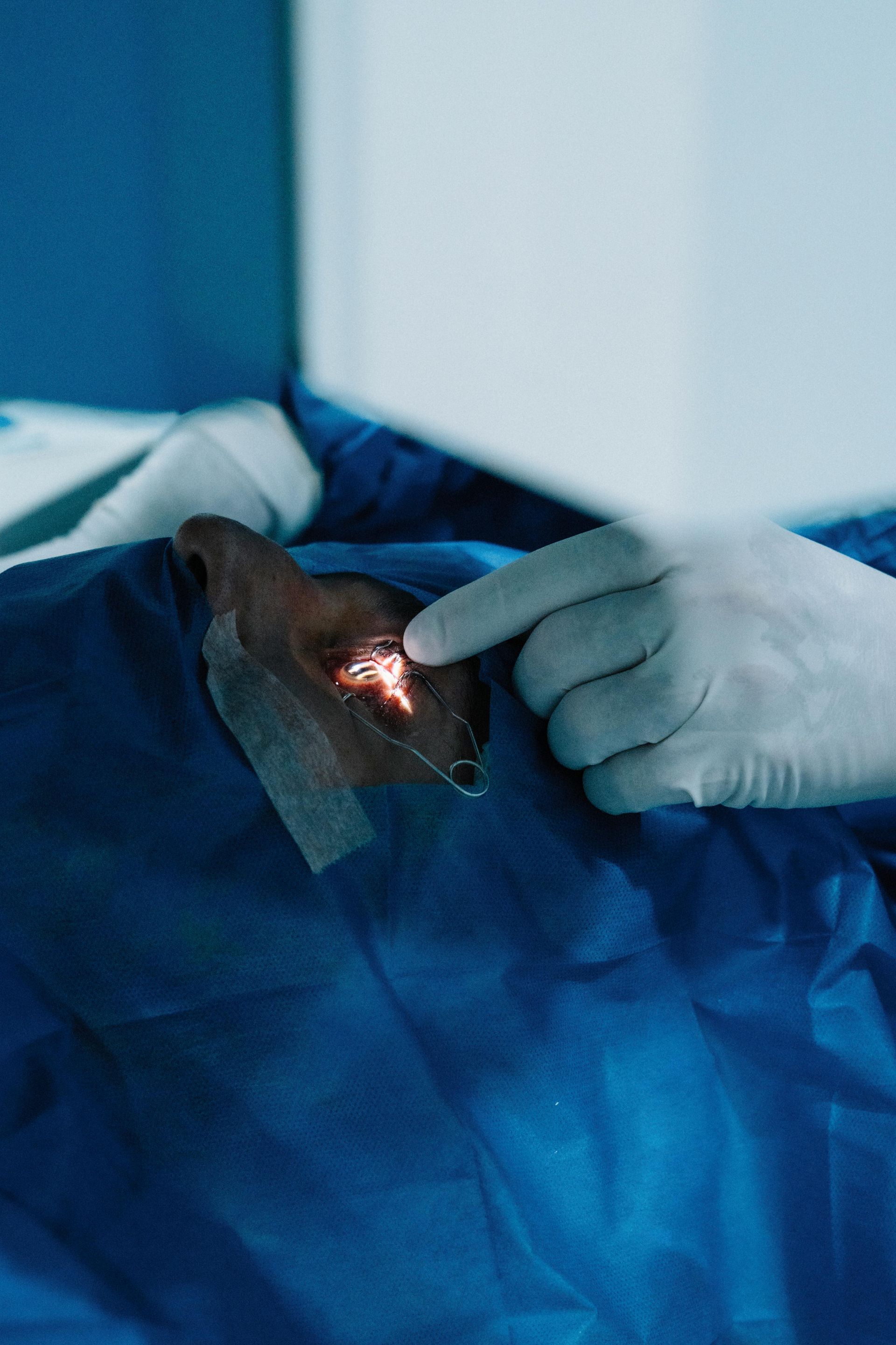
At Burlington Eyecare, we see patients that take hydroxychloroquine, which is a less toxic version of chloroquine, when under treatment for RA. Over the long haul, the drug can be toxic to the macula and neurons in the eye, causing thinning and atrophy of the ganglion cells and a condition called “bull’s eye maculopathy”. The toxicity can result in loss of acuity, colour vision defects, and central visual field loss; damage which is, unfortunately, irreversible.
The key is to monitor regularly, catch it early, and then jig around dosages and treatments to optimize comfort and minimize damage. This is for the rheumatologist to sort out; we see the patient annually for a complete eye exam. About 7.5% of chloroquine patients will have retinal damage: the longer the exposure, the greater incidence of retinopathy.
It was great to read the September issue of BMC Ophthalmology (read here). Early detection is critical. However, macular appearance is hard to detect. Identifying central visual field loss is important, but it shows up later than retinal thinning. The studies in this article were done with optical coherence tomography (OCT) and the researchers found that the ganglion cell complex (GCC), which is quite thick and multilayered at the macula, starts thinning and is affected first in toxicity.
We have been doing OCT’s on all our patients for years and can catch abnormalities very early on. The protocol for detecting toxicity in the eye has been acuities, macular biomicroscopy, and central field testing. Baseline OCT’s should now be at the top of this list.
It is great to have the tools to detect damage and other problems early. The OCT allows us to look at the actual tissues at the back of the eye, which is a really wonderful thing.
‘til next week,
The good doctor