Cataract surgery: the patient’s experience

Blog vol 5.17. Cataract surgery: the patient’s experience.


Last Thursday, one of my colleagues at Burlington Eyecare underwent a procedure that you do twice in your life. No, it was not having your second child or your second hip replacement. It was cataract surgery. Nowadays, almost all people will have the procedure done, one eye at a time. It can be scary, like most surgeries there are risks involved, and when it comes to your eyes and your sight, that is daunting.


The eye doctor will sometimes mention that the cataract is “ripe”. What they mean is that it is now worth the “trouble” of replacing it with an implant lens. It is important to note that this is surgery and it is a serious matter. There is an optimal window of time to have cataracts removed, because the opacity in the eye will become more and more brittle over time, making its removal really tricky.


Fortunately, we have very capable surgeons and hospital staff to refer to in our area.


The process started months ago.  My colleague was referred for cataract assessment, and it was determined that the procedure would be beneficial. Next was lens power determination, where the focal length of the eye was measured with a special machine to determine the power of the implant lenses needed. The type of implant lens was decided upon, then the custom lenses were ordered and set aside for the big day. They went with a mono-focal lens to correct distance vision, and upgraded to an aspheric lens to help with night vision.


The worst eye is usually done first (unless visual impairment is present in the worst eye) and in this case, the right eye was done on Thursday. They arrived at the Hamilton Regional Eye Institute which is on the King Campus of St. Joseph’s Healthcare around 11:00 am and were admitted and taken to the Eye Centre where preparation included gel drops and a small IV to enable the administration of fluids and a sedative. Onto the gurney and into the OR, however, after this not much is remembered. During the surgery, light mostly and the whirring of a machine (a phaco machine, a machine used in cataract surgeries).


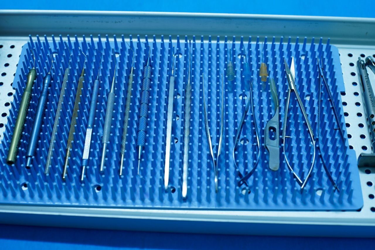
While the sedation was working, the surgeon prepped the area and made a small incision, about 3 mm in size, and through that port and a widely dilated iris, cut through the front of the capsule, removed it and then with ultrasound and suction broke down the cataract and removed it, careful at all times to not damage the capsule of the lens (the sac) where the lens implant will be centred. The anterior chamber was filled with Healon to assist implantation, then the folded implant lens was inserted through the port and into the capsule, unfolded and centered, and finally the surgeon exited the port and ensured that the eye’s pressure would secure the wound. Pretty brilliant really.  (Read more about the procedure here).


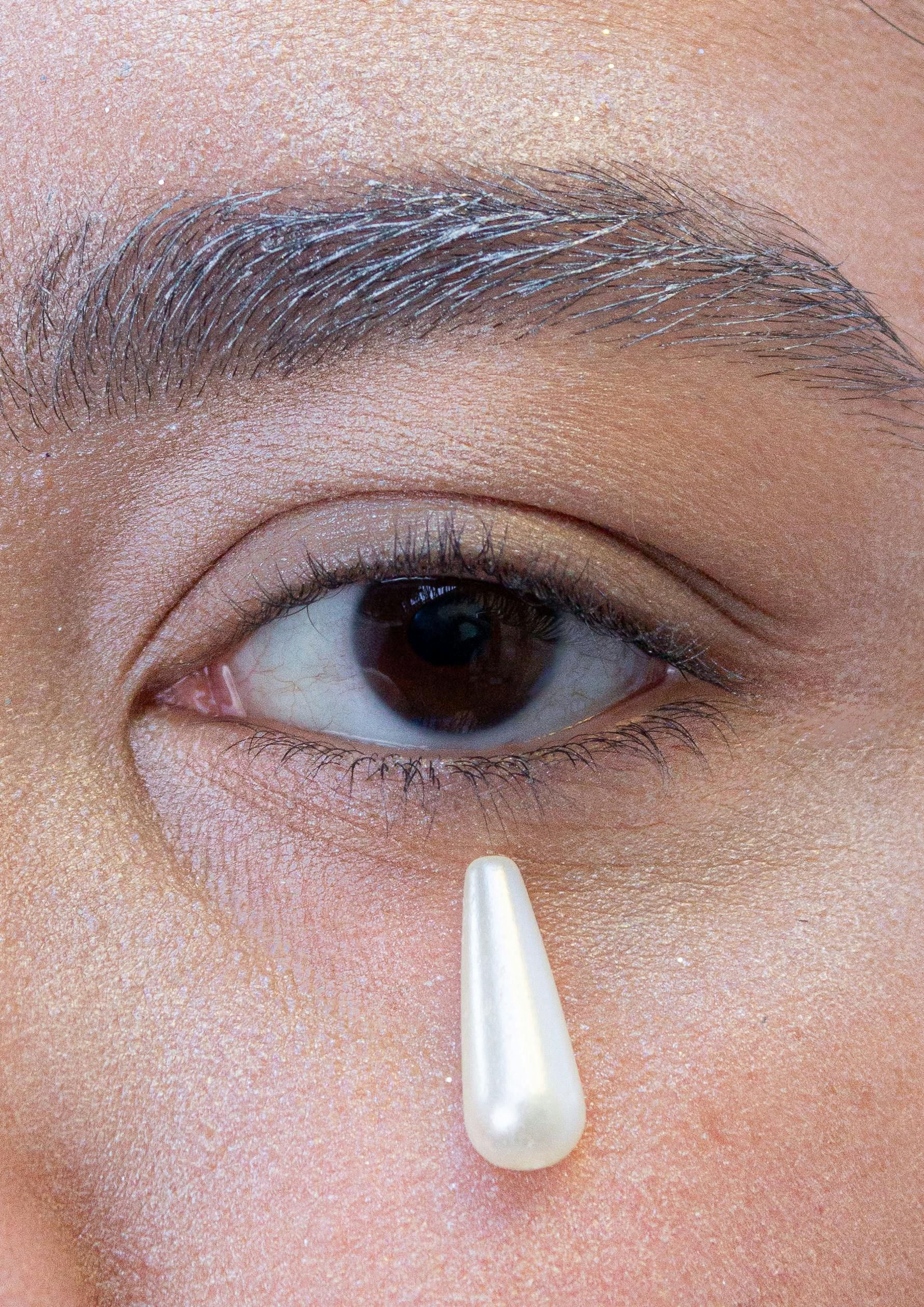
This all took 10-20 minutes, no glitches. My colleague was soon home and able to remove the eye patch. First experience: “Everything is so bright!” The cataract eye is like an incandescent light and the post-surgery eye is like LED, or high definition, which at first is dazzling. The eye looks great, a bit of redness and swelling, but healing nicely. The “patient” is using steroid drops and finishing a round of antibiotics with lots of lubrication drops, and is back to work.


The second eye is scheduled in the next few weeks, then wow! so bright, so clear. 


The good doctor


By Dr. Mark Germain May 1, 2026
The good doctor discusses how the eye is a window to the heart with the help of the OCT.
The good doctor take a deeper look at how gut health is connected to eye tissues.
By Dr. Mark Germain April 29, 2026
Blog vol 6.40. The Gut and the Eyes. One of my biggest jobs is to keep abreast of new research and advances in eye health. Most recently, we have seen new stem cell research, gene therapies, as well as new drugs for dry and wet macular degeneration. In January of this year, there was an article in the journal, Cell Death and Disease, that looked at cell apoptosis, specifically with the photoreceptors of the eye. (Read the article here ). Apoptosis is the genetic programming of deliberate and constructive cell death, allowing for the maintenance of healthy structures and systems in the body. Some estimates have the body replacing 50 to 70 billion cells daily by this process. Necrosis, on the other hand, is unplanned cell death caused by injury, toxins, or other negative outside factors; this causes inflammation and disrupts the body’s balance of things. There are stressors that speed up cell turnover. The study looked at photoreceptors in the retina and were able to demonstrate cellular recovery, even in late stages of apoptosis, recovery not breakdown. When certain stressors were reduced, by working with the mitochondria, there was recovery of the photoreceptor cells. How? Mitophagy, the mitochondria’s own process of programmed breaking down and then repurposing the recycled components for new ones. This mitochondrial repair helps to reverse the sequence of apoptosis for the photoreceptors. Mitochondria, how cool. How is this helpful for our eyes? Conditions like retinitis pigmentosa or macular degeneration directly involve the photoreceptors and may be slowed or reversed by helping the mitochondria. So, let’s take care of those mitochondria. There is some evidence for nutritional support for the mitochondrial system. The nutrient, Urolithin A (UA), which is a compound made in our gut from fiber found in pomegranates, berries, and nuts is the only clinically proven substance known to trigger mitophagy, made in the gut no less. Vitamin B3 is critical for mitochondria function, however vitamin B3 based supplements are still to be clinically tested for their support of cell recovery. Nutrients Coenzyme Q10 and L-carnitine are known to help the mitochondria generate energy, and interestingly, infrared light therapy has also been shown to benefit mitochondrial function. Again, food for thought. The gut and its function needs a lot of research. How do probiotics and nutrient precursors affect eye tissues? It is so interesting that the gut can be a factor in eye health. Maybe adding Urolithin A as a supplement is wise, along with the antioxidants in Lutein and Zeaxanthin we use to combat photoreceptor degeneration. Will keep you posted on any new developments.  The good doctor
By Dr. Mark Germain April 16, 2026
The good doctor focuses in on visual processing. Why do we miss things that are right in front of our eyes?
By Dr. Mark Germain April 10, 2026
The good doctor discusses the Artemis II mission, space travels affects on the eyes, and "the dark side of the moon"
By Dr. Mark Germain April 3, 2026
The good doctor discusses exciting new and emerging options to treat nystagmus.
By Dr. Mark Germain March 27, 2026
The good doctor reflects on The Country of the Blind, a book by Andrew Leland which gives insight into the experience of vision loss.
More Posts